Phase I to Phase IV Clinical Trials
Become part of one of our clinical trials
DermCare Experts, through its affiliated site Beacon Clinical Research, conducts Phase I to IV clinical trials for a variety of skin diseases. Our staff have extensive experience in managing trials for leading pharmaceutical companies worldwide. We have a complete research unit with all necessary equipment and certification. The work at Beacon Clinical Research additionally spun off Clinical Research IO, www.clinicalresearch.io, now considered one of the top three electronic trial management systems in the industry.
DermCare Experts is equipped
with the following research equipment:
- Bed, shower and kitchen facilities
- Dedicated research exam rooms
- Secure drug storage room
- Locked cabinet for controlled substances
- Wireless Internet
- Internet-based video security
- Fully equipped laboratory
- 2-8 Celsius refrigerator
- -20 freezer
- -80 freezer
- Dry ice
- Uninterrupted power supply
Clinical Trials
In addition to the therapies currently on the market, there are more treatment options undergoing efficacy and safety assessments through clinical trials. We have a robust clinical research arm in our affiliated research site that conducts Phase II-IV trials on atopic dermatitis, psoriasis, and actinic keratosis.
Learn more about these conditions below and if you are interested in participating in our clinical trials, please contact us at info@dermcare.expert.
 Atopic dermatitis, or eczema, presents as dry skin, itch, and rash.
Who does it affect?
It is the most common inflammatory skin disease worldwide, affecting about 230 million people. It often manifests as part of the “atopic triad” which includes atopic dermatitis, asthma, and hay fever, with food allergies also included in the cluster of “atopic tendencies”. Atopy, or this tendency to develop an over-active immune response to environmental factors, is mostly inherited. Atopic dermatitis affects 20% of children, often starting in infancy and usually developed before the age of 6. However, people of all ages can develop this condition.
What causes atopic dermatitis?
There is no single cause of atopic dermatitis, as it results from a complex combination of environmental and genetic factors including the body’s immune system, skin structural gene mutations, skin cell defects, and the skin microbiome. The current literature identifies atopic dermatitis to be primarily an immune-mediated inflammatory response regulated by cytokines. These are small proteins produced by cells that control the growth and activity of other immune system cells and blood cells. The primary cytokines involved from current research are IL-4, IL-13, and IL-22. The over-production of these cytokines cause a defect in the skin barrier and inflammation that manifest in the features seen in atopic dermatitis patients.
Atopic dermatitis is a systemic disease manifesting in the skin, so events that disrupt the body’s normal balance in other ways may trigger flares. These events can range from a viral infection or eating certain foods that the body is allergic to, or may not be obvious at all. Given atopic dermatitis presents as dry skin, environmental factors that make the skin even drier may make the symptoms harder to control. These include the winter weather, frequent washing particularly with very hot water, soaps and harsh detergents, and low humidity in the air.
How is it treated?
Treatment options vary widely depending on the severity of your condition, your work environment, and your lifestyle. At DermCare Experts, we are committed to finding the best treatment plan tailored to every patient after understanding and assessing your individual needs. We treat atopic dermatitis with the most up-to-date knowledge and access to the most recent therapies as well as cutting-edge clinical trials which pay you for your time and effort.
General measures can help with mild cases of atopic dermatitis and prevent flares:
- Avoid skin irritants wherever possible. This includes harsh soaps and detergents, abrasive washcloths in the shower, coarse clothing fabrics, chemicals and antiseptics, perfumes and cosmetics.
- Keep the skin hydrated at all times with emollients, or moisturizers. Emollients should be applied to the skin when it is still wet
- Antihistamines such as Zyrtec or Allegra to provide relief for itch
Topical treatments can resolve symptoms of itch and inflammation during flares:
- Topical corticosteroids (e.g. betamethasone or clobetasol) to reduce inflammation of the skin
- Topical immunomodulating agents for low-term maintenance of recurrent flares, especially in sensitive areas such as the face, skin folds, or genitals
- Topical calcineurin inhibitors (e.g. tacrolimus or pimecrolimus)
- Topical PDE4 inhibitors Eucrisa® (crisaborole)
- Topical JAK inhibitor Opzelura® (ruxolitinib), approved by FDA July 2022
Systemic treatments can treat moderate-to-severe cases inadequately controlled with topicals:
- Biologics are among the most advanced available today for atopic dermatitis. They are synthesized from living organisms and target very specific protein receptors. Both of the following are administered by subcutaneous injections
- Dupixent® (dupilumab) blocks IL-4 and IL-13
- Adbry® (trakolinumab-ldrm) blocks IL-13
- Oral JAK inhibitors
- Cibinqo® (abrocitinib)
- Rinvoq® (upadacitinib)
- Traditional systemic drugs that suppress the immune system (e.g. methotrexate, cyclosporine, mycophenolate mofetil, azathioprine)
- Phototherapy, most commonly with narrow-band UVB
Different Types of Psoriasis
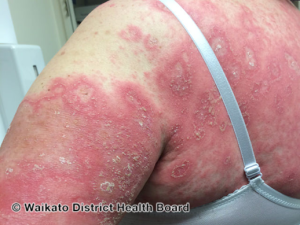
Psoriasis generally presents as scaly thickened areas of the skin. However, there are different types of psoriasis affecting various areas of the body including the scalp, nails, palms, and soles with different presentations. Psoriasis can be localized or generalized, which differ in treatment rationale.
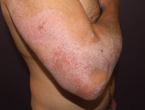
Plaque Psoriasis
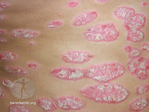 The most common type is plaque psoriasis that presents as well-demarcated plaques of thickened, scaly skin. It most likely affects elbows, knees, and lower back but may arise on any part of the body. Plaque psoriasis can also affect the palms and soles, referred to as palmoplantar psoriasis. This can occur as localized palmoplantar psoriasis or as part of generalized psoriasis.
Inverse Psoriasis
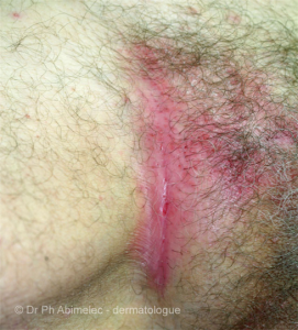 Inverse psoriasis, sometimes referred to as flexural psoriasis, is psoriasis localized to the skin folds (e.g. armpits, groin, under breasts) and genitals. Instead of the scale usually associated with plaque psoriasis, it tends to present as shiny and smooth. There may be a crack in the depth of the skin crease.
Nail Psoriasis
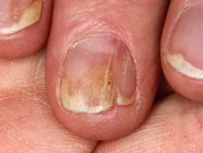 Nail psoriasis is commonly associated with plaque psoriasis, affecting 90% of patients with chronic plaque psoriasis at some point in their lifetime. It is characterized by pitting, onycholysis, yellowing, and ridging of the fingernails and/or toenails.
Guttate Psoriasis
Guttate psoriasis presents as small “raindrops” of red, scaly skin, hence the name guttate deriving from “gutta” meaning drop. It is a form of acute psoriasis and usually clears within 3-4 months. However, persistent guttate psoriasis may develop into chronic plaque psoriasis.
Pustular Psoriasis
 Pustular psoriasis is a more rare type of psoriasis, and is most. It causes reddish, scaly, pus-filled bumps. Patients who have generalized pustular psoriasis should seek immediate attention as widespread pustular psoriasis can be life-threatening.
What causes psoriasis?
Psoriasis is an immune-mediated inflammatory disease. It is a multifactorial condition with different factors contributing to its presentation including genetics, comorbidities such as obesity and metabolic syndrome, and lifestyle choices such as smoking and stress. The pathways leading to psoriasis and its clinical presentation are still being explored, but it is clear that immune factors and cytokines are responsible for the clinical features of psoriasis. Currently, it is known that IL1β and TNFα are two cytokines involved. Theories are also exploring the roles of IL17A and the TH17 pathway in psoriasis. Because of the inflammatory and immune-mediated response associated with psoriasis, patients with psoriasis often have psoriatic arthritis in conjunction.
There are additional triggers for the various types of psoriasis. Guttate psoriasis often develops 1-2 weeks after a streptococcal infection of the upper respiratory tract, particularly tonsillitis. It has also been reported to develop after COVID-19 infection. General pustular psoriasis can occur in pregnant women triggered by changes in hormone, calcium, and elafin levels on a predisposing genetic background.
How is psoriasis treated?
Treatment options vary widely depending on the severity of your condition, your work environment, and your lifestyle. At DermCare Experts, we are committed to finding the best treatment plan tailored to every patient after understanding and assessing your individual needs. We treat atopic dermatitis with the most up-to-date knowledge and access to the most recent therapies as well as cutting-edge clinical trials which pay you for your time and effort.
Over-the-counter medications can be used to treat mild cases of psoriasis and maintain clearance
- Emollients are recommended for all psoriasis patients to prevent painful cracking and scaling
- Keratolytic agents such as urea or salicylic acid is recommended for palmoplantar psoriasis to thin down the thick scaling skin
- Coal tar has anti-inflammatory and anti-scaling properties to thin down thick scale. This is especially useful for scalp psoriasis to allow medicated shampoo and other topical medications reach the scalp.
Prescription topical medications are also be used to treat mild and localized psoriasis
- Topical steroids which vary in strength (e.g. mometasone, betamethasone, clobetasol) to reduce inflammation and cut down on itch
- Vitamin-D like compounds (calcitriol and calcipotriene) as the first line of therapy and for maintenance
- Calcineurin inhibitors (ie. tacrolimus or pimecrolimus) especially in sensitive areas like the face or skin folds where long-term use of topical corticosteroids is not indicated
- New topical medications are available that targets new;y discovered pathways
- Roflumilast (Zoryve®) newly approved by FDA in July 2022 that is a PDE4 inhibitor
- Tapinarof (Vtama®) newly approved by FDA May 2022 that is an AhR modulating agent
Systemic treatments to treat generalized psoriasis that is inadequately controlled with topicals
- Biologics are among the most advanced available today for atopic dermatitis. They are synthesized from living organisms and target very specific protein receptors. These medications are administered as a subcutaneous injection
- TNFα inhibitors: etanercept (Enbrel®), adalimumab (Humira®), and certolizumab (Cimzia®)
- IL-17 agents: secukinumub (Cosentyx®), ixekizumab (Taltz®), and brodalumab (Siliq®)
- IL-12 and IL-23: ustekinumab (Stelara®), guselkumab (Tremfya®), and risankizumab (Skyrizi®)
- Oral medication apremilast (Otezla®), a PDE4 inhibitor
- Oral medication deucravacitinib (Sotyktu®) newly approved by the FDA in September 2022, a small molecule compound that targets tyrosine kinase 2
- Traditional systemic medications that suppress the immune system (ie. methotrexate, ciclosporin)
- Oral retinoid acitretin (Soriatane®)
- Phototherapy with narrowband UVB or a combination of psoralen and UVA
Most of the treatments above are indicated for plaque psoriasis. There are some treatments available that are indicated only for other types of psoriasis
- Spesolimab-sbzo (Spevigo®) newly approved as the first approved treatment for generalized pustular psoriasis flares in adults
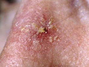 Actinic keratosis, or solar keratosis, is a precancerous lesion caused by UV damage to the skin.
Who does it affect?
Actinic keratoses can affect virtually anyone; however, certain risk factors may increase the chance of developing these precancerous lesions. These risk factors include, but are not limited to, having an extensive history of sun exposure or sunburns, living in a sunny environment (tropics, subtropics, etc.), working outside, and having a weakened immune system.
What causes actinic keratoses?
The main cause of actinic keratosis lesions is oftentimes frequent or intense sun exposure either through natural UV rays or tanning beds. These lesions are slow-growing and are considered to be precancerous because of their potential to develop into a squamous cell carcinoma. If caught early, there are a variety of different treatments that can help stop this progression.
How is it treated?
There are a multitude of different treatment options including cryotherapy, field therapy, excision or shave, electrodessication and curettage. Each of these treatment options are explained in further detail below.
Field treatments target visible and developing sunspots on a large area (ex. face, scalp, or both) of the body. There are different modalities used for field treatment including photodynamic therapy (PDT), imiquimod cream, or fluorouracil 5% cream.
- Photodynamic therapy is also known as blue light therapy and is mainly used for treatment of the face, ears, neck and scalp. The procedure starts with applying aminolevulinic acid gel. After absorbance of the gel, the treated areas are exposed to blue light for approximately 1,000 seconds (16 minutes and 40 seconds). The aminolevulinic acid gel makes actinic keratoses extremely sensitive to light. Therefore, any sun-damaged cells would be destroyed during the process.
- Fluorouracil 5% cream (5-FU) is a cytotoxic topical treatment for these precancerous lesions. This cream is prescribed in combination with calcipotriene 0.005% cream, which enhances the effectiveness of the treatment and decreases the treatment duration. The combination cream treatment is commonly prescribed twice daily for 2 to 6 weeks depending on the target area and prescriber discretion.
After undergoing treatment many patients will experience mild to exuberant reactions at the target areas, pictured below.
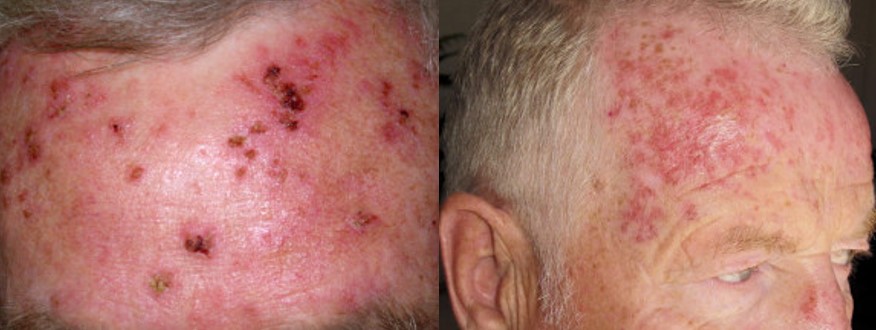
- Imiquimod cream is an immune response modifier topical treatment often used for clearance of actinic keratoses. Similar to fluorouracil 5% cream, this treatment modality is quite successful in decreasing reoccurrence of actinic keratoses at treated areas. This cream is frequently used three times a week over the span of three to four months.
Cryotherapy is another approach for destroying these types of lesions. This process includes spraying liquid nitrogen for 5 to 20 seconds on the point of concern. Predominantly, this treatment is favored when treating individual, visual actinic keratosis lesions. Liquid nitrogen treated points will blister, swell, and appear red. However, these symptoms often subside after approximately 7-10 days.
Excisions can be performed on actinic keratoses but leave a permanent scar. This treatment ensures that the entire lesion has been removed using a surgical blade under local anesthesia. This treatment is primarily preferred when there is an isolated lesion of concern. After the procedure, the specimen diagnosis will be confirmed with pathology.
Lastly, shaving, electrodessication and curettage can be performed on singular actinic keratosis lesions. This is a small in-clinic procedure that includes shaving of the lesion, curettage (scrapping) at the base of the lesion and electrodessication (burning). Oftentimes, the healing process after this operation is much longer and most likely will leave a permanent scar.